![]() Figure 5 of
Macnamara, Mol Vis 2004;
10:51-56.
Figure 5 of
Macnamara, Mol Vis 2004;
10:51-56.
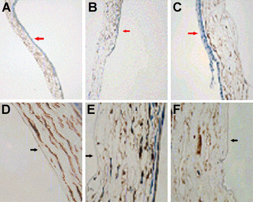
Figure 5. Aquaporin-1 expression in normal versus injured mouse corneas
Representative photographs of aquaporin-1 expression in uninjured mouse corneas (A,D), 1 day after hot sterile water injection (B,E), and 7 days after hot sterile water injection (C,E). The magnification of A, B, and C was 20x and the magnification of D, E, and F was 100x. In A, B, and C the red arrows identify the epithelium. In D, E, and F the black arrows identify the endothelium.