![]() Figure 1 of
Macnamara, Mol Vis 2004;
10:51-56.
Figure 1 of
Macnamara, Mol Vis 2004;
10:51-56.
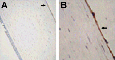
Figure 1. Aquaporin-1 expression in normal human cornea
Representative photographs of aquaporin-1 expression in normal human corneal specimens. The magnification of A was 20x and the magnification of B was 100x. Arrows identify the endothelium.