![]() Figure 6 of
de Iongh, Mol Vis 2004;
10:566-576.
Figure 6 of
de Iongh, Mol Vis 2004;
10:566-576.
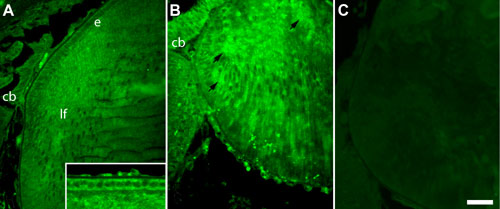
Figure 6. Expression of ALK3 protein in transgenic lenses
Immunofluorescence of ALK3 in sections of P2 FVB (A) and OVE550 (B, C) mouse lenses. A: In the wild type FVB lenses at P1 distinct reactivity for ALK3 was detected in the anterior lens epithelium (e) and in the cortical fibers (lf). Inset shows higher magnification view of cytoplasmic reactivity for ALK3 detected in the anterior epithelial cells. Reactivity was also present in the ciliary body (cb). B: OVE550 lenses there was distinctly increased reactivity for ALK3 in the fiber cells (arrows) and in the ciliary body (cb). C: No specific reactivity was detected with normal goat serum. The scale bar in C represents 75 μm (A-C) or 25 μm (inset).
