![]() Figure 5 of
de Iongh, Mol Vis 2004;
10:566-576.
Figure 5 of
de Iongh, Mol Vis 2004;
10:566-576.
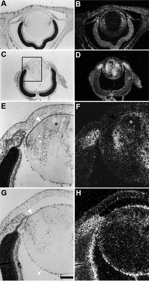
Figure 5. Expression of ALK3 and BMPRII mRNA during aberrant lens differentiation
In situ hybridizations of ALK3 (B,D,F) and BMPRII (H) on sections of P1 OVE591 (A,B) and OVE550 (C-H) mouse lenses. Bright field views (A,C,E,G) of hematoxylin stained sections are shown for orientation. In phenotypically normal transgenic (OVE591) lenses at P1, aberrant expression of ALK3 was detected in the cortical fibers (B; arrowheads). Expression in the lens epithelium, cornea and retina and ciliary body/iris were similar to wild type expression. In transgenic mice, which express higher levels of transgene (OVE550) and have developed a phenotype by P1 (C) the aberrant expression of ALK3 in the fibers was stronger (D). Increased expression of ALK3 was also detected in the ciliary margins of the optic cup. At higher magnification (E,F) expression for ALK3 is apparent in fibers that are starting to show nuclear pyknosis (arrowheads) but not in fibers that have degenerated (asterisk). There appeared to be no aberrant expression of BMPRII in lens fibers of OVE550 mice (H). The peripheral region of the lens epithelium appeared to have reduced levels of BMPRII expression (H; arrowhead) whereas the central region tended to exhibit stronger levels of expression. As in wild type lenses, distinct expression was present in the tunica vasculosa lentis (H; arrow). The scale bar in G represents 500 μm (A-D) or 150 μm (E-H).