![]() Figure 3 of
de Iongh, Mol Vis 2004;
10:566-576.
Figure 3 of
de Iongh, Mol Vis 2004;
10:566-576.
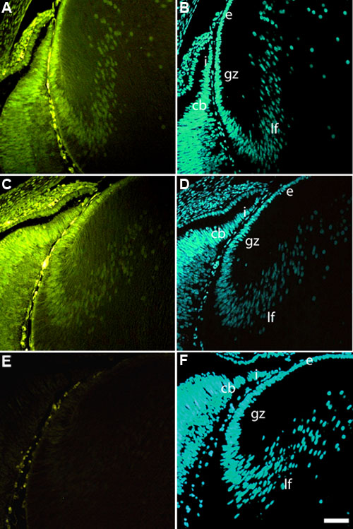
Figure 3. Localization of phosphorylated Smads in lens
Embryonic day 18 rat lenses were immuno-stained with antibodies for phospho-Smad1 (A), phospho-Smad2 (C) or non-immune rabbit IgG (E) and counterstained with Hoechst (bisbenzimide) nuclear dye (B,D,F). Immunofluorescence for phospho-Smad1 (A) and Smad2 (C) was detectable in the nuclei of the lens epithelium (e) and fibers (lf) as well as ciliary body (cb) and iris (i). Strongest reactivity appeared to be in the germinative zone (gz) of the epithelium. No reactivity was detected with the non-immune rabbit IgG (E). The scale bar represents 100 μm.