![]() Figure 2 of
de Iongh, Mol Vis 2004;
10:566-576.
Figure 2 of
de Iongh, Mol Vis 2004;
10:566-576.
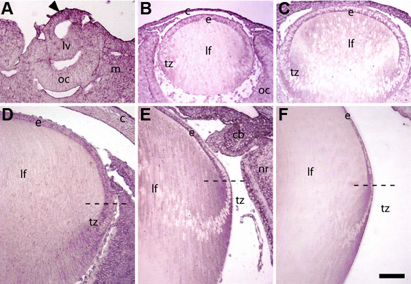
Figure 2. Expression of ActRIIA mRNA during lens differentiation
In situ hybridization of ActRIIA on sections of E12 (A), E14 (B), E16 (C), E18 (D), P3 (E), and P21 (F) rat eyes, using digoxigenin labeled riboprobes. At E12 (A), ActRIIA was expressed strongly in anterior lens vesicle (lv) cells (arrowhead). Weaker expression was detected in the posterior lens vesicle (lv), and peri-ocular mesenchyme (m). From E14 to P21 (B-F) distinct expression was present in the lens epithelium (e), including the transitional zone (tz) but decreased in the lens fibers (lf). Transcripts were also detectable in the optic cup and cornea during embryonic development. In postnatal eyes at P3 (E) strong expression was detected in the ciliary body and the ganglion cell and neuroblast layers of the neural retina (nr). The scale bar in F represents 100 μm (A-E) or 250 μm (F).