![]() Figure 1 of
de Iongh, Mol Vis 2004;
10:566-576.
Figure 1 of
de Iongh, Mol Vis 2004;
10:566-576.
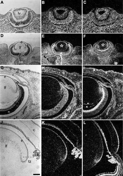
Figure 1. Expression of ALK3 and BMPRII mRNA during lens differentiation
Expression of BMP receptors was examined by in situ hybridization of sections of E12 (A-C), E14 (D-F), E18 (G-I), and P21 (J-L) rat eyes, using specific radiolabeled riboprobes for ALK3 (B,E,H,K) and BMPRII (C,F,I,L). Sections stained with hematoxylin (A,D,G,J) show ocular structure at each stage. At early lens vesicle stage (E12), ALK3 (B) and BMPRII (C) were expressed ubiquitously in the lens vesicle (lv), optic cup (oc) and extraocular mesenchyme. In the late lens vesicle (E14), ALK3 (E) and BMPRII (F) transcripts were detected in lens epithelium (e) but decreased in the primary fibers (lf), particularly those in the center of the lens that had reached the greatest degree of elongation (asterisks). Transcripts for both receptors were detected in the optic cup (oc) and extraocular mesenchyme (m). ALK3 was detected in the optic nerve (on) and BMPRII along the inner optic cup (oc). In E18 lenses, distinct ALK3 expression (H) was detected in lens epithelium (e) but decreased in differentiated fibers (lf). Transcripts were also detected throughout the neural retina (nr), in cornea (c), ciliary and iridial retina (cb) and eyelids (ey). BMPRII (I) was similarly detected in lens epithelium and decreased in fibers. It was expressed strongly in the ganglion cell (g) layer, but weakly in the neuroblast (n) layer, of the neural retina. Weaker expression was detected in the ciliary and iridial retina, epidermis of eyelid and extraocular mesenchyme (m). Expression was also found in the tunica vasculosa lentis (arrowheads). In lenses of weanling animals (J-L), ALK3 (K) and BMPRII (L) transcripts were detectable in the lens epithelium (e) including the transitional zone (tz). Little or no expression was detected in the mature fibers (lf). Strong expression was also detected in the ciliary body, iris and corneal epithelium and endothelium. Distinct BMPRII expression is also visible in layers of the neural retina. The dashed line in J indicates the equator where epithelial cells commence elongation into fiber cells. The scale bar in J represents 100 μm (A-C) or 250 μm (D-L).