![]() Figure 2 of
Corti, Mol Vis 2004;
10:533-536.
Figure 2 of
Corti, Mol Vis 2004;
10:533-536.
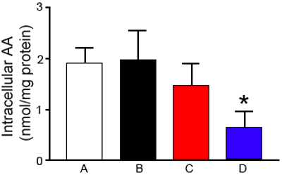
Figure 2. Effect of UV light on intracellular ascorbic acid
Cell monolayers were incubated for 15 min in Dulbecco's phosphate buffered solution containing 1 g/l glucose in the presence of 0.5 mmol/l dehydroascorbic acid. After washing, the monolayers were incubated in fresh Dulbecco's phosphate buffered solution containing glucose alone. Some were treated with 0.3 mmol/l 1,3-bis(chloroethyl)-1-nitrosourea and/or exposed to the UV source (exposure to the UV source was always the last step). Column A is the control and was incubated with dehydroascorbic acid (DHAA) only. Column B was incubated with DHAA and exposed to UV. Column C was incubated with DHAA and then incubated with 1,3-bis(chloroethyl)-1-nitrosourea. Column D was incubated with DHAA, then treated with 1,3-bis(chloroethyl)-1-nitrosourea and exposed to the UV source. At the end of the incubations ascorbic acid content of cell monolayers was determined and expressed as nmol/mg protein. Values presented are the mean of three separate experiments; the error bars represent the standard deviation. The asterisk above bar "D" indicates the value was significantly different from C (p=0.028).
