![]() Figure 1 of
Corti, Mol Vis 2004;
10:533-536.
Figure 1 of
Corti, Mol Vis 2004;
10:533-536.
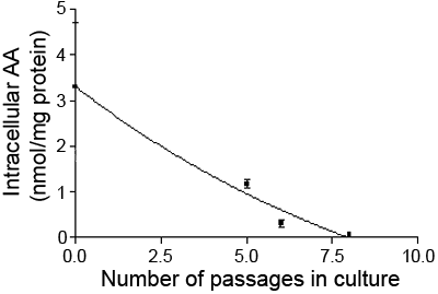
Figure 1. Decline of the content of ascorbic acid during culture of bovine lens epithelial cells
Cell monolayers were maintained in RPMI 1640 medium as described in "methods", and the content of ascorbic acid assessed at passages 0, 5, 6, and 8; the ascorbic acid content of the original explant fragment at plating into the culture dish is reported as passage 0. Each point represents the mean of three independent determinations; the error bars represent the standard deviation. The best fit curve is shown.
