![]() Figure 6 of
Mansfield, Mol Vis 2004;
10:521-532.
Figure 6 of
Mansfield, Mol Vis 2004;
10:521-532.
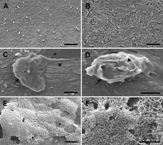
Figure 6. Morphological changes in lens epithelial explants cultured with TGFβ and FGF
Explants were cultured without TGFβ (A,B) or with TGFβ (C-F) for 1 day then without FGF (A) or with FGF (B-F). TGFβ2 was added at a final concentration of 100 pg/ml (C,D) or 50 pg/ml (E,F) and FGF-2 at 100 ng/ml (B-D) or 20 ng/ml (E,F). Explants were cultured for 21 days (A-D) or 30 days (E,F) with change of medium every 5 days and re-addition of FGF as before (B-F only), before fixing and processing for SEM. Explants cultured without addition of growth factors retained the cobblestone morphology typical of the normal lens epithelium (A). A corresponding explant cultured with FGF alone showed elongation of cells consistent with FGF induced fiber differentiation (B), whereas a TGFβ/FGF treated explant cultured in parallel exhibited formation of small plaques (C,D, arrows) and spindle-like cells (C, arrowhead). Although this explant was generally well covered with cells, a region of denuded capsule can be seen (C, asterisk). The surface of one plaque was relatively smooth (C), whereas the surface of a nearby plaque (D) was irregular with small, globular protrusions (D, arrowhead). A TGFβ/FGF treated explant from another experiment is shown in E. In this explant, cells retracted into thick mounds during extended culture leaving large regions of the lens capsule denuded (E, asterisk). At higher magnification, the cellular surface of this explant was seen to be covered with a thick web of extracellular matrix-like material (F). The bar represents 50 μm in A,B, 20 μm in C,D, 100 μm in E, and 10 μm in F.
