![]() Figure 3 of
Mansfield, Mol Vis 2004;
10:521-532.
Figure 3 of
Mansfield, Mol Vis 2004;
10:521-532.
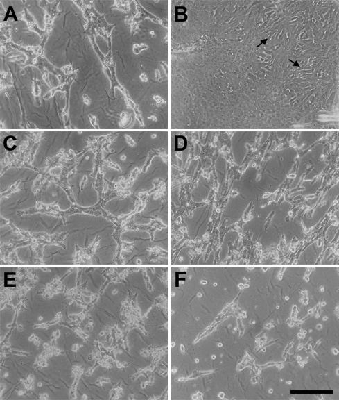
Figure 3. Effects of FGF and other growth factors on TGFβ induced cell loss
Lens epithelial explants were cultured with 50 pg/ml TGFβ2 for 1 day, then medium was replaced and explants were cultured without further additions (A) or with immediate addition of FGF (B), EGF (C), PDGF (D), IGF (E), or HGF (F). Growth factors were used at the following final concentrations: FGF-2, EGF and PDGF at 10 ng/ml, IGF at 50 ng/ml, and HGF at 20 ng/ml. The explants were photographed four days later. Addition of FGF, but not the other growth factors, prevented the cells from undergoing typical TGFβ induced changes that lead to cell death and loss from the explant. In explants treated with TGFβ alone (A) or TGFβ plus growth factors other than FGF (C-F), the wrinkled denuded lens capsule was visible between rafts of abnormal cells and scattered dying cells, whereas the TGFβ/FGF treated explant (B) remained covered with cells, including some spindle-like cells (arrows). The bar represents 180 μm.