![]() Figure 2 of
Mansfield, Mol Vis 2004;
10:521-532.
Figure 2 of
Mansfield, Mol Vis 2004;
10:521-532.
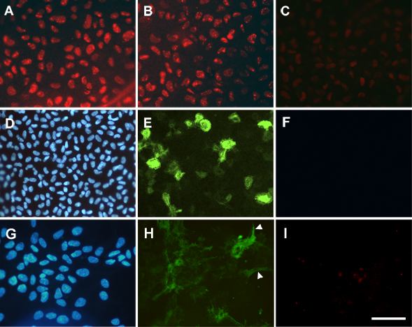
Figure 2. Immunolocalization of Pax6 and αSMA in whole mounts
Explants were cultured without growth factors (A) or with 100 ng/ml FGF-2 (B,C) for 3 days (A,B) or 8 days (C), or precultured for 1 day with 25 pg/ml TGFβ2 followed by 5 ng/ml FGF-2 and a further 4 days of culture (D-F), or for 2 days with 75 pg/ml TGFβ without addition of FGF (G-I). Immunolocalization of Pax6 (A-C,F,I) and αSMA (E,H) was carried out by a double labeling technique with Hoechst counterstaining of nuclei (D,G). D-F and G-I, each row represents images of the same region. Strong nuclear reactivity for Pax6 was detected in explants cultured with a high, fiber differentiating dose of FGF on day 3 (B), as in no growth factor controls (A), but reactivity was much weaker on day 8 (C). In TGFβ/FGF treated explants, although only low concentrations of each growth factor were used in this experiment, Pax6 reactivity was lost within 5 days of culture (F), while scattered cells expressed αSMA (E). An explant fixed at an early stage of TGFβ induced transdifferentation is depicted in G-I. While many cells were beginning to express αSMA, few exhibited the morphology characteristic of more mature spindle cells (indicated by arrowheads in H). Nevertheless, virtually all nuclear reactivity for Pax6 had already been lost (I). The bar represents 60 μm in A-I.