![]() Figure 1 of
Mansfield, Mol Vis 2004;
10:521-532.
Figure 1 of
Mansfield, Mol Vis 2004;
10:521-532.
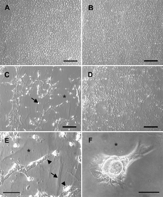
Figure 1. The effect of FGF on TGFβ treated lens epithelial explants
Explants were cultured without TGFβ (A,B) or with TGFβ (C-F) for 1 day. Medium was then replaced without further addition of TGFβ and FGF was added to some cultures (B,D,F). TGFβ2 was used at a final concentration of 25 pg/ml (C-E) or 50 pg/ml (F), FGF-2 at 5 ng/ml (B,D) or 20 ng/ml (F). Explants were photographed after a total of 5 days of culture using Integrated Modulation Contrast (A-E) or phase contrast (F). In the absence of growth factors (A), the cells remained in the cobblestone array typical of the normal lens epithelium. FGF alone (B), added on day 1, had no apparent effect on cellular morphology. Addition of TGFβ alone (C,E) induced elongation of cells and extensive cell loss, exposing large regions of denuded lens capsule (asterisk), while many bright, rounded, dying cells were visible (C, arrow). At higher magnification (E), cell surface blebbing (arrowheads) and wrinkling of the lens capsule (arrow) were also evident. Generally, addition of FGF to TGFβ pretreated explants prevented these morphological changes and resulted in a marked reduction in the loss of cells from the explant (D). In the explant depicted, the capsule was almost completely covered with healthy cells. In the TGFβ/FGF treated explant shown in F, which was poorly covered with cells at the start of the experiment and received higher doses of growth factors, an isolated patch of monolayered cells piled up into a thick plaque during culture, exposing the underlying lens capsule (asterisk). The bar represents 90 μm.