![]() Figure 3 of
Unsoeld, Mol Vis 2004;
10:468-475.
Figure 3 of
Unsoeld, Mol Vis 2004;
10:468-475.
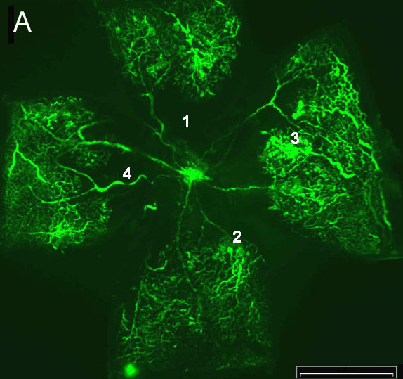
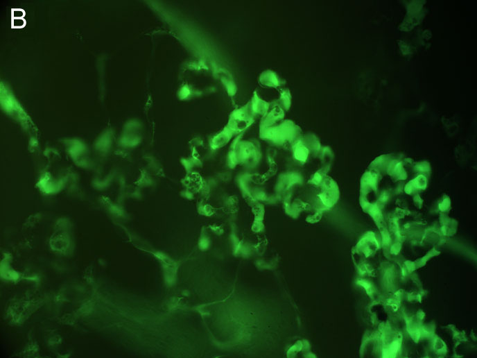
Figure 3. Flatmount of a mouse retina with oxygen-induced retinopathy
A: Ischemic retina from a 17 day old mouse that had been exposed to hyperoxia from days 7 to 12 of life (x20). Numbers indicate the typical features of proliferative retinopathy: (1) central avascular area, (2) blood vessel tufts, (3) presumed extra-retinal neovascularization, (4) blood vessel tortuosity. B: Magnification of A showing a cluster of retinal proliferations (presumed extra-retinal neovascularization; x80). Scale bar represents 1000 μm.