![]() Figure 2 of
Unsoeld, Mol Vis 2004;
10:468-475.
Figure 2 of
Unsoeld, Mol Vis 2004;
10:468-475.
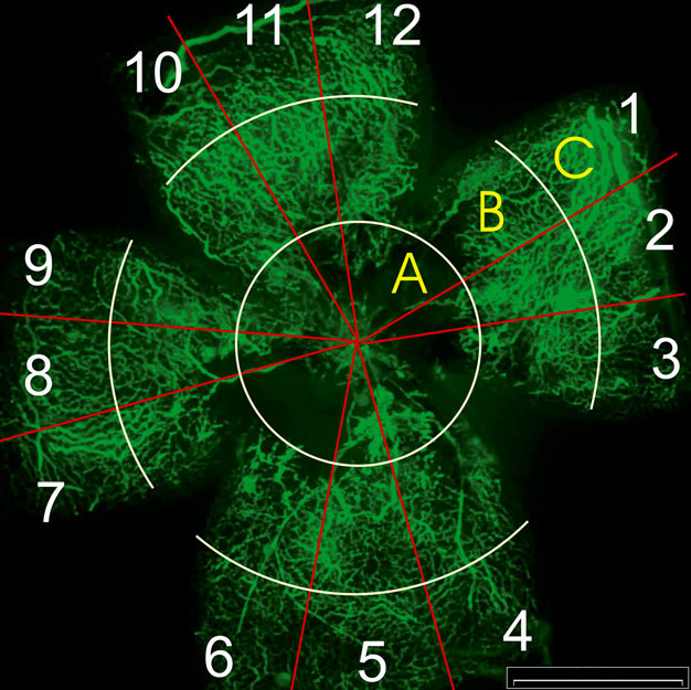
Figure 2. Retinal flat mount with superimposed zones and "clock hours"
A retinal flat mount after perfusion. The retina was divided into 3 concentric zones: the inner zone around the optic disc (A), the middle zone (B), and the outer zone (C) for the scoring of the central avascular area. Vascular proliferations were quantified by counting blood vessel tufts and presumed extra-retinal neovascularization in each of 12 equally sized sections of the retina (numbers). Scale bar represents 1000 μm.