![]() Figure 1 of
Leske, Mol Vis 2004;
10:43-50.
Figure 1 of
Leske, Mol Vis 2004;
10:43-50.
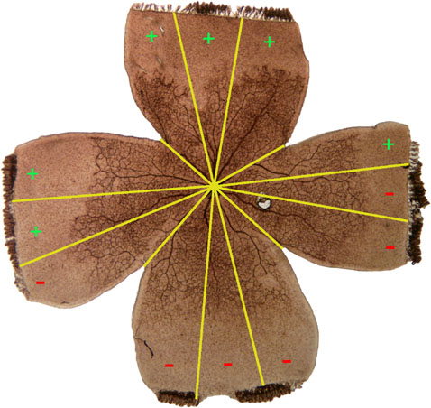
Figure 1. Neovascularization grading method
Each quadrant of flat-mounted retinae was divided visually into three equal parts for a total of 12 sectors (clock hours) and examined by light microscopy for the presence or absence of neovascularization. Clock hours with neovascularization present were graded positive. All clock hours graded positive were summed to give the total neovascularization score (0-12 clock hours) for the retina. This retina received a score of 6 clock hours.