![]() Figure 3 of
Stitt, Mol Vis 2004;
10:432-438.
Figure 3 of
Stitt, Mol Vis 2004;
10:432-438.
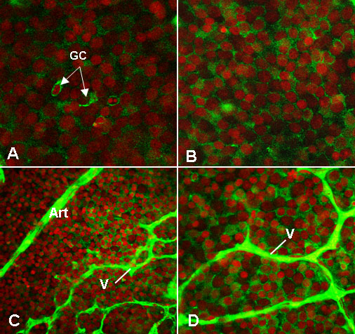
Figure 3. PEDF immunolocalization in retinal flatmounts in OIR mice at P13
Optical sectioning of retinal flatmounts from a non-punctured eye reveals distinct PEDF immunoreactivity in the cytolplasm of the retinal ganglion cells (GC; A). This PEDF staining appears more widespread and intense in the punctured eye (B). In ganglion cells adjacent to veins/venules (v) in punctured eyes (C and D) there is intense PEDF immunoreactivity. This intensity appears reduced in ganglion cells adjacent to arterioles (Art). Original magnification for A, B, and D is x200; for C is x100.