![]() Figure 2 of
Stitt, Mol Vis 2004;
10:432-438.
Figure 2 of
Stitt, Mol Vis 2004;
10:432-438.
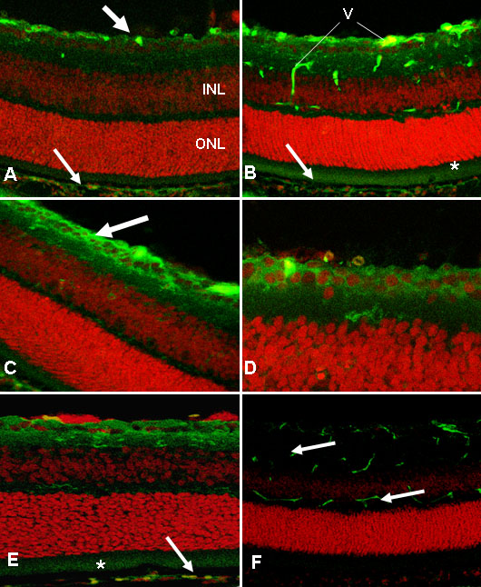
Figure 2. PEDF immunolocalization in retina from OIR mice: alteration by ocular perforating injury
A: Confocal scanning laser micrograph of a retinal cryosection from a P13 non-punctured eye. There is distinct PEDF-immunoreactivity (green) in the ganglion cell layer (arrow). Propidium iodide (PI) stain (red) shows the retinal layers (INL, inner nuclear layer; ONL, outer nuclear layer). There is some staining in the RPE (arrow). B: In the P17 non-punctured eye, the retina also shows PEDF in the ganglion cells with staining in the RPE (arrow) and interphotoreceptor matrix (indicated by an asterisk ["*"]). Vascular labelling is evident (V) as the anti-mouse secondary antibody detects IgGs in the blood column (arrows). C: In the punctured eye at P13, there is more intense PEDF-immunoreactivity in the ganglion cell layer (arrow; compare with A). D: Higher magnification of a P13 retina following perforating injury shows PEDF immunolocalization in the cytoplasm of the ganglion cells. E: By P17 in the perforated eye, PEDF immunoreactivity in the retina is diminished in comparison to P13, but there is comparable staining in the RPE (arrow) and interphotoreceptor matrix (indicated by an asterisk ["*"]). F: Primary antibody omission control shows the vascular labelling (arrows). A, B, C, D, and F original magnification x100. E original magnification x200.