![]() Figure 1 of
Stitt, Mol Vis 2004;
10:432-438.
Figure 1 of
Stitt, Mol Vis 2004;
10:432-438.
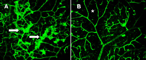
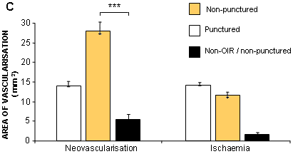
Figure 1. Penetrating ocular injury inhibits pre-retinal neovascularization in oxygen induced retinopathy (OIR)
A and B: Confocal scanning laser micrographs of P17 retinas in which the microvasculature has been visualized using lectin labeling of endothelial cells. A: Retina from non-punctured (control) eye in which fronds of pre-retinal vessels are evident (arrows). B: In comparison to A, the retina from the punctured eye shows less neovascularization, although large areas of retinal ischaemia (as evidenced by areas of non-perfusion) are still evident; an asterisk ("*") marks one such area. Original magnifications x40. C: Quantification of pre-retinal neovascularization and areas of non-perfusion (classified as ischaemic regions) in normal (non-hyperoxia-exposed) and OIR mice (un-punctured and punctured). It is apparent that puncture wounds to the eye have a significant anti-angiogenic effect (triple asterisks ("***") indicate p<0.001) when compared to non-punctured controls, although retinal ischaemia remains unaffected.