![]() Figure 6 of
Fautsch, Mol Vis 2004;
10:417-425.
Figure 6 of
Fautsch, Mol Vis 2004;
10:417-425.
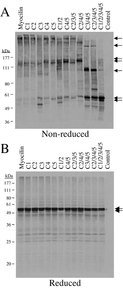
Figure 6. Disulfide bond formation in mutagenized myocilin
A: 35S-labeled proteins obtained from in vitro transcription/translation reactions (40 μl) were immunoprecipitated with anti-hemagglutinin antibody. Immunoprecipitated products were bound to protein A-sepharose, washed in RIPA buffer, and separated on a 7% SDS-PAGE gel under denatured, non-reduced conditions. Gels were transferred to PVDF membrane followed by autoradiography. B: 35S-labeled proteins obtained from in vitro transcription/translation reactions (10 μl, not immunoprecipitated) from each construct were separated on 11% denatured, reduced SDS-PAGE gels. Gels were transferred to PVDF membrane followed by autoradiography. Canine microsomal membranes were added to in vitro transcription/translation reactions. Arrows indicate prominent myocilin complexes. Asterisk represents the top of the gel.