![]() Figure 3 of
Smith, Mol Vis 2004;
10:392-398.
Figure 3 of
Smith, Mol Vis 2004;
10:392-398.
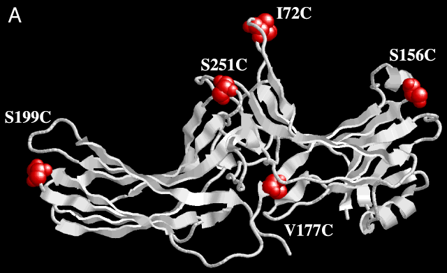
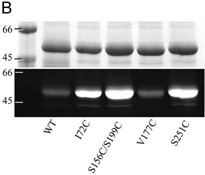
Figure 3. Fluorescent labeling of cysteine-substituted arrestin
A: Three dimensional representation of the crystal structure of arrestin, indicating the amino acids (space filled in red) for which cysteines were substituted. B: Purified arrestin (native, I72C, S156C/S199C, V177C, and S251C) was reacted with AlexaFluor-594 maleimide and separated on 12% SDS PAGE. Proteins were stained with coomassie brilliant blue (upper bands), or excited with ultraviolet light (lower bands).