![]() Figure 3 of
Wahlin, Mol Vis 2004;
10:366-375.
Figure 3 of
Wahlin, Mol Vis 2004;
10:366-375.
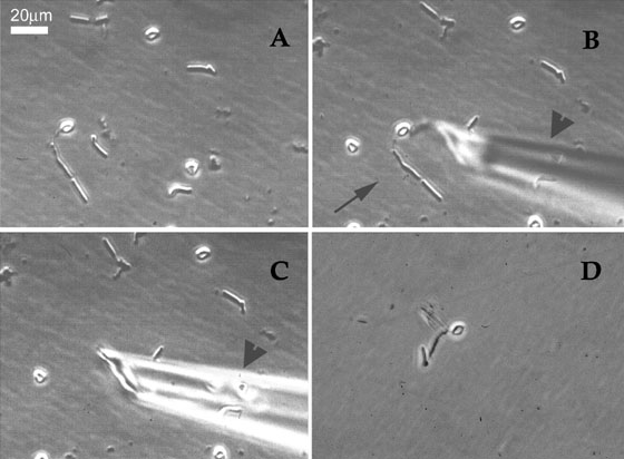
Figure 3. Capture of single cells for cDNA synthesis
A: Retinal dissociates contained a mixture of cells with defined morphologies, round cells, and cell debris. B: shows a rod photoreceptor cell (arrow) being approached by a thin microcapillary pipette (arrowhead) which is lowered with a micromanipulator. C: The rod photoreceptor cell has been aspirated into the capillary micropipette through the application of slight negative pressure (arrowhead). D: The cell captured in C has been released into a fresh, buffer-containing dish, from which it will be recaptured with a fresh micropipette.