![]() Figure 2 of
Wahlin, Mol Vis 2004;
10:366-375.
Figure 2 of
Wahlin, Mol Vis 2004;
10:366-375.
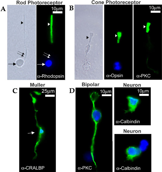
Figure 2. Immunocytochemical characterization of dissociated cells from adult mouse retina
Primary antibody binding was detected in all cases with secondary antibodies labeled with Alexafluor-488, which fluoresces green. DAPI-stained cell nuclei were visualized in the blue channel. A: Rod photoreceptor cell shown by phase contrast microscopy (left) and fluorescence microscopy (right). Opsin immunoreactivity is exclusively detected in the cell's outer segment (arrowhead). The inner segment (double arrow) is devoid of fluorescence; the cell nucleus is indicated with an arrow. B: Cone photoreceptor cells visualized by phase contrast microscopy (left panel) and by immunofluorescence with an anti-cone opsin antibody (middle panel) and with an anti-PKC antibody (right panel), which stains a subpopulation of cones. Both opsin and PKC immunoreactivities are localized to the cone outer segment (arrowheads). C: Müller glial cell immunoreacted with an antibody against cellular retinaldehyde binding protein (CRALBP). The nucleus is localized near the center of the cell (arrow); CRALBP immunoreactive materials are detected throughout the cell, including its processes. D: Neuron-like cells immunoreacted with antibodies against PKC (left panel) and calbindin (right panel). The PKC-positive cell displayed on the left has a morphology consistent with a bipolar identity, quite different from the PKC-positive cone cell shown in B. The calbindin-positive cells on the right lack characteristic neuronal features, but their immunoreactivity suggests that they could be either horizontal or amacrine cells.