![]() Figure 1 of
Wahlin, Mol Vis 2004;
10:366-375.
Figure 1 of
Wahlin, Mol Vis 2004;
10:366-375.
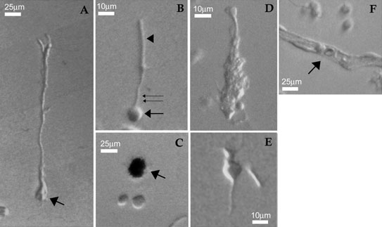
Figure 1. Morphological characterization of dissociated cells from adult mouse retinas
A: Müller glial cell; arrow indicates putative end feet. B: Rod photoreceptor cell with a round cell body (arrow), inner segment (double arrow) and outer segment (arrowhead). C: Heavily pigmented retinal epithelial cell (arrow). Two process free cells are also displayed in this panel. D,E: Putative neuronal cells whose identity is difficult to determine; the cell in D appeared to have several retracted processes. F: Small capillary type blood vessel, containing a red blood cell (arrow).