![]() Figure 2 of
Koizumi, Mol Vis 2004;
10:328-340.
Figure 2 of
Koizumi, Mol Vis 2004;
10:328-340.
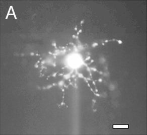
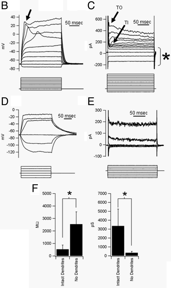
Figure 2. IIR in amacrine cell dendrites
Amacrine cells were recorded in a horizontal slice preparation and then injected with Lucifer yellow. A: Example of an amacrine cell with single slow action potentials, injected with Lucifer yellow at the end of recording. Scale bar, 10 μm. B: Current-clamp experiment. Injected currents were from -20 pA to 160 pA, in 20 pA steps, for 200 msec. Resting membrane potential was -69 mV. This cell generated single slow action potentials (arrow), but their thresholds were very high (around -30 mV). C: Voltage-clamp experiment. Voltages were clamped from -91 mV to +19 mV, in 10 mV steps, for 200 msec. Arrow with TI, transient inward current. Arrow with TO, transient outward current. Asterisk, hyperpolarization-induced IIR. D: IIR is not observed in amacrine cells whose dendrites are removed in the process of preparation. Current-clamp experiment. Injected currents were from -40 pA to +60 pA, in 20 pA steps, for 200 msec. E: Voltage-clamp experiment. Voltages were clamped from -111 mV to -41 mV, in 10 mV steps, for 200 msec. F: Comparison between amacrine cells with intact dendrites (n=33) and those without dendrites (n=7). Amacrine cells without dendrites have higher input resistance (2545±992 MΩ, n=7) than those with dendrites (526±351 MΩ, n=33). Resting membrane conductance in amacrine cells without dendrites (328±173 pS, n=7) were lower than in those with dendrites (3356±1893 pS, n=33). *Significant difference by student t-test, p<0.001.