![]() Figure 1 of
Koizumi, Mol Vis 2004;
10:328-340.
Figure 1 of
Koizumi, Mol Vis 2004;
10:328-340.
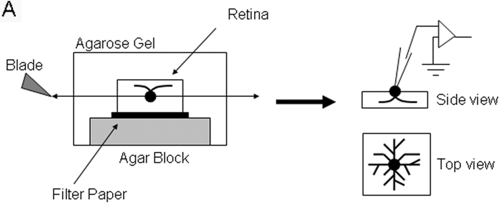
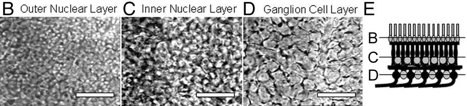
Figure 1. Horizontal slice preparation of the mouse retina
The retina was prepared and sliced horizontally as shown in the schematic and images. A: The retina was isolated from the eyecup and attached to filter paper, which was flattened on an agar block and covered by low-temperature melting agarose gel. The retina was cut at the level of the inner nuclear layer with a vibratome. The slice was mounted in a recording chamber and whole-cell patch-clamp recordings were made. In this preparation, the dendrites of amacrine cells were well preserved, and the whole dendritic field could be visualized with Lucifer yellow. B-E: Views of the horizontally sliced retina at different levels. All scale bars represent 50 μm.