![]() Figure 2 of
Cullinan, Mol Vis 2004;
10:315-322.
Figure 2 of
Cullinan, Mol Vis 2004;
10:315-322.
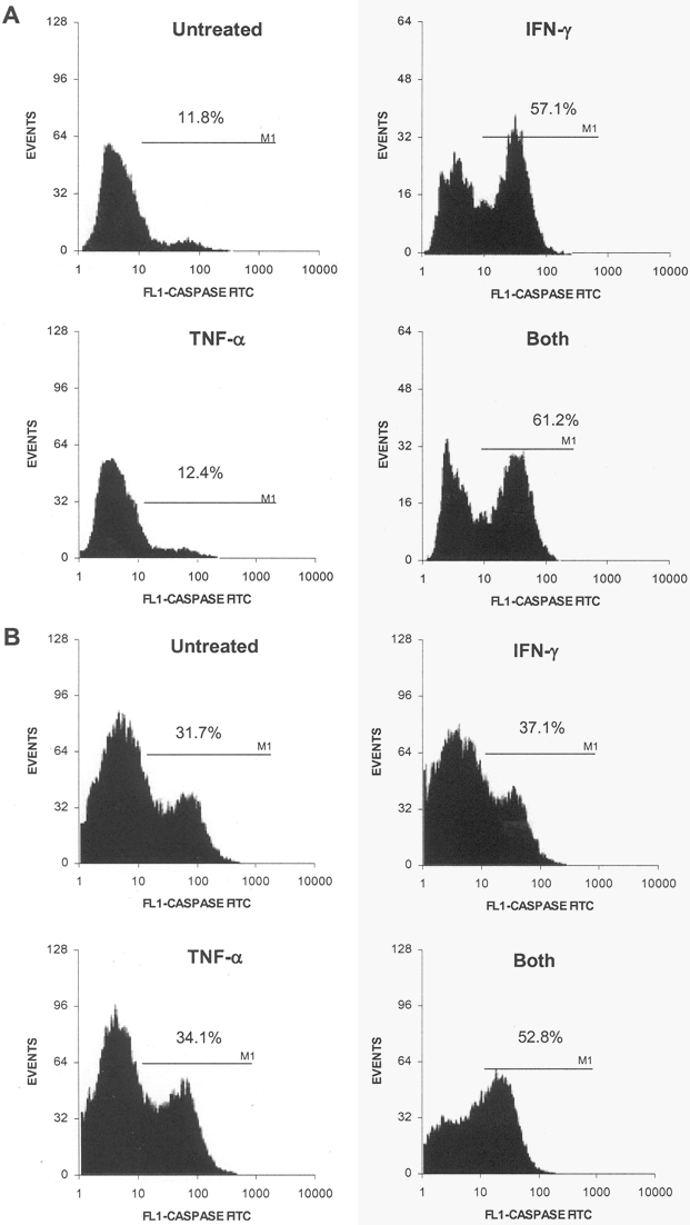
Figure 2. Caspase 3 activation in normal and cytokine-treated Y79 and Weri Rb-1 cells
Caspase-3 cleavage was measured in cytokine-treated Y79 (A) or Weri Rb-1 cells (B). A total of 1x10⁶ Y79 or Weri Rb-1 cells were treated with 50 ng/ml of recombinant human IFN-γ, TNF-α, or both cytokines (50 ng/ml of each) for 72 h in medium containing 10% FBS. The cells were then fixed, permeabilized, stained with an antibody-specific for the activated form of caspase-3, and quantitated by FACS. The bars represent the area used to calculate the percentage of cells positive for active caspase-3.