![]() Figure 1 of
Cullinan, Mol Vis 2004;
10:315-322.
Figure 1 of
Cullinan, Mol Vis 2004;
10:315-322.
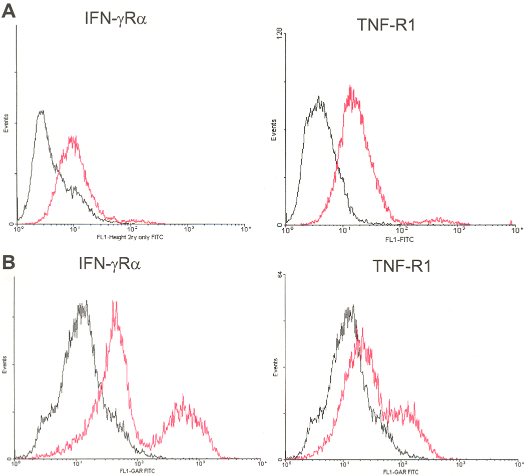
Figure 1. The presence of IFN-γ and TNF-α receptors on retinoblastoma cells
A total of 1x10⁶ Y79 (A) or Weri Rb-1 (B) cells cultured in medium containing 10% FBS, were stained with polyclonal antibodies specific for the IFN-γRα, the TNF-R1, or with a non-specific rabbit IgG control antibody. The cells were then fixed, washed as described in Materials and Methods, and stored on ice until they were quantitated by FACS analysis. The black line represents the signal with control non-specific IgG and the red line represents the signal with receptor specific antibody.