![]() Figure 4 of
Mukhopadhyay, Mol Vis 2004;
10:304-314.
Figure 4 of
Mukhopadhyay, Mol Vis 2004;
10:304-314.
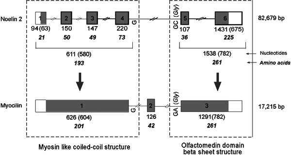
Figure 4. Comparison of gene structure of Noelin 2 and Myocilin
The sizes of the exons of both the genes and the number of amino acids encoded by those are shown. Exons of Noelin 2 used in noelin 2_v1 and predicted to have been fused in Myocilin during evolution are indicated. The total number of nucleotides within these sets of exons for the genes, number of nucleotides in the coding sequence (in parenthesis), and number of amino acids coded by the exon(s) are shown. The codon split by intron 4 of Noelin 2 and the terminal bases for the codons at the 3' end of exon 1 and 5' end of exon 3 of Myocilin are indicated. Five of the six exons of Noelin 2 sharing similar features with other 2 Noelin genes are represented by bolded square. The protein domains encoded by Noelin 2 and myocilin are marked.