![]() Figure 3 of
Mukhopadhyay, Mol Vis 2004;
10:304-314.
Figure 3 of
Mukhopadhyay, Mol Vis 2004;
10:304-314.
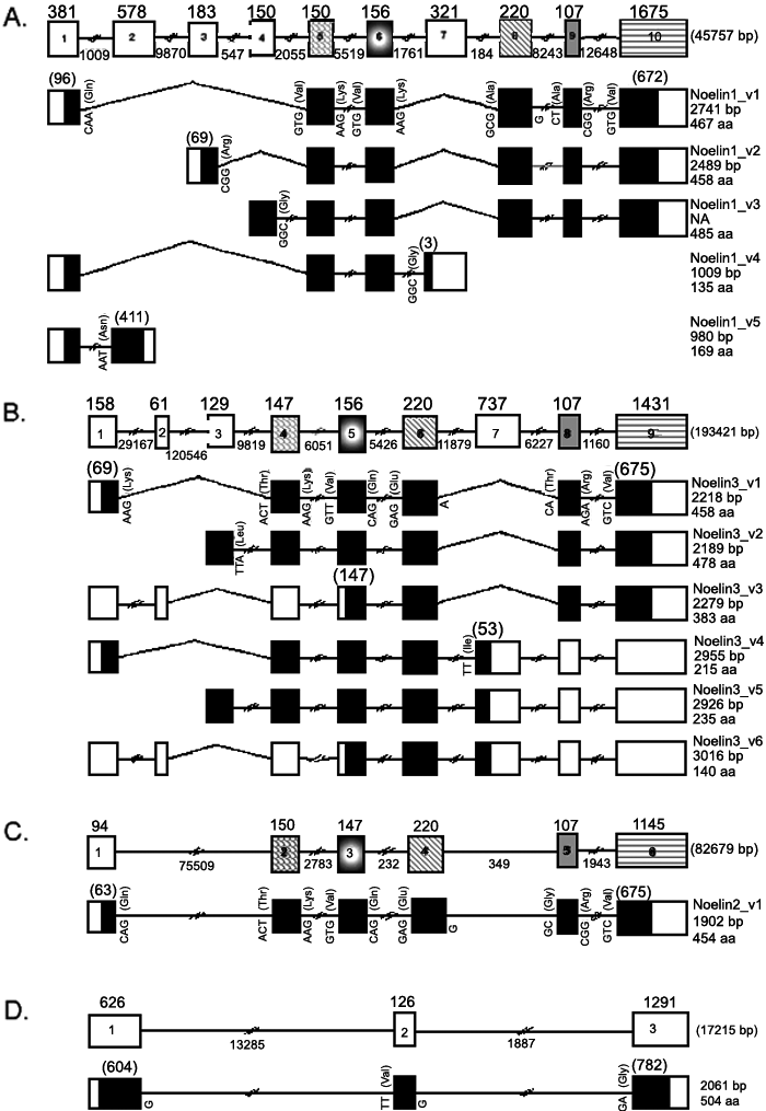
Figure 3. Structure of "myocilin related" genes and alternate usage of exons
Sizes of exons and introns of Noelin 1 (A), Noelin 3 (B), Noelin 2 (C) and Myocilin (D) genes are shown at the top of each panel. Exons are numbered from the 5' to 3' direction. Usage of different exons in the splice variants is shown below the structures of the respective genes. The black and white areas within the exons denote coding sequences and untranslated regions (UTRs), respectively. The number of nucleotides used for coding amino acids in any exon that also contains an UTR has been given in parenthesis above the exon-symbol. Exons not completely characterized at the 5' end are shown by broken symbols (e.g., exon 4 of Noelin 1 and exon 3 of Noelin 3). Complete or partial sequence of the terminal triplet codons of the exons at splice junctions and the corresponding amino acids are shown. For example, where intron sequence in the gene is present within a triplet codon, the nucleotides of the codon as distributed between adjacent exons are shown. All three Noelins contain 5 common exons (exon numbers 5, 6, and 8-10 in Noelin 1; 4-6 and 8-9 in Noelin 3; and 2-6 in Noelin 2) which are indicated by different patterns. Each of these 5 exons (denoted by a specific pattern) is similar in size and/or the terminal codons in all 3 Noelins. The sizes of the genes, cDNA of the splice variants of each gene and the number of amino acids in the corresponding isoforms are shown, as appropriate, in the right side of the illustration. The names of the different isoforms have been given according to the recommendations of Human Genome Organization.