![]() Figure 4 of
Brown, Mol Vis 2004;
10:281-288.
Figure 4 of
Brown, Mol Vis 2004;
10:281-288.
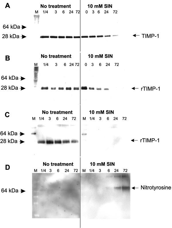
Figure 4. SIN-1 degrades both TIMP-1 and recombinant TIMP-1 (rTIMP-1)
A: Aliquots of culture media from normal cell cultures were collected, SIN-1 was added for varying time periods, and then western blot analysis with monoclonal TIMP-1 antibody to the carboxyl terminal region was performed. Note that the untreated samples showed stable TIMP-1 levels. In contrast, the SIN-1 treated serum-free media showed less staining of the TIMP-1 protein over time. B: Aliquots of recombinant TIMP-1 (rTIMP-1, 50 ng) were exposed to SIN-1 for varying time periods and then analyzed by western blotting with theTIMP-1 antibody to the carboxyl terminal region. Note that the rTIMP-1 protein in the untreated samples remained stable over time. The rTIMP-1 in the SIN-1 treated samples showed decreased staining by 24 h and was not seen by the 72 h time point. C: Aliquots of recombinant TIMP-1 (rTIMP-1, 50 ng) were exposed to SIN-1 for varying time periods and then analyzed by western blotting with theTIMP-1 antibody to the loop 1 site of the TIMP-1 molecule. The untreated rTIMP-1 samples were stable over time. The SIN-1 treated rTIMP-1 samples did not stain at all even at the 15 min time period. D: The blot from C was stripped and reprobed with an antibody to nitrotyrosine. No bands were seen in the untreated samples. After 24 and 72 h treatment with SIN-1, an approximately 68 kDa band was seen.