![]() Figure 3 of
Brown, Mol Vis 2004;
10:281-288.
Figure 3 of
Brown, Mol Vis 2004;
10:281-288.
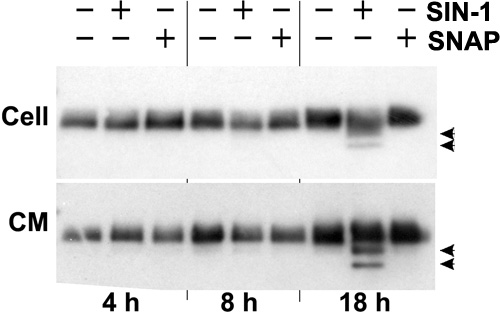
Figure 3. TIMP-1 protein is degraded after SIN-1 treatment of cell cultures
This is a representative western blot to identify TIMP-1 protein in normal cell cultures after incubation with or without 1 mM SIN-1 or 1 mM SNAP. Samples were separated into two fractions, the cell lysate (Cell) and culture media (CM). The TIMP-1 band migrates at 28 kDa. Note that in the 18 h SIN-1 treated samples, there were lower molecular weight bands, representing TIMP-1 fragments (arrowheads).