![]() Figure 4 of
Loewen, Mol Vis 2004;
10:272-280.
Figure 4 of
Loewen, Mol Vis 2004;
10:272-280.
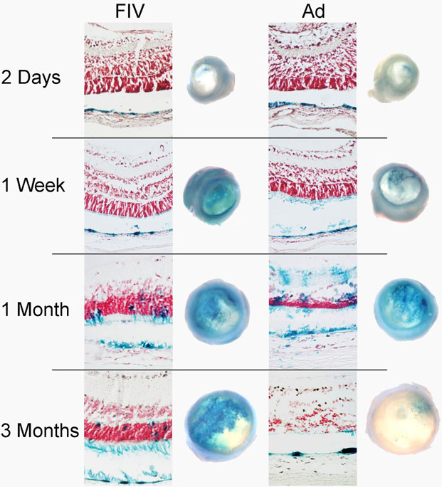
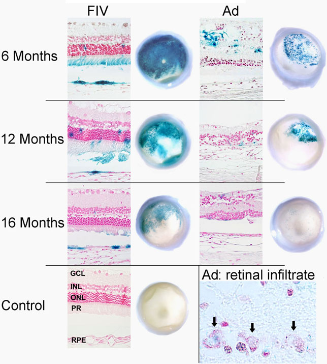
Figure 4. Representative eye cups and histological sections
Representative eye cups and histological sections through the transduced retina of eye pairs from the same animal. Right eyes were injected with FIV, left eyes with Ad vector. When a retinal detachment was present in the section, images of neurosensory retina and RPE are shown in their normal spatial relationship. Insert: large, macrophage-like cells with abundant cytoplasm appeared to contain phagocytosed β-galactosidase positive material (arrows). Macrophage infiltrates were often seen in Ad transduced eyes after 3 months. Abbreviations used in the "control eye" section are: GCL (retinal ganglion cell layer), INL (inner nuclear cell layer), ONL (outer nuclear layer), PR (photoreceptor outer segments), RPE (retinal pigment epithelium).