![]() Figure 4 of
Pina, Mol Vis 2004;
10:265-271.
Figure 4 of
Pina, Mol Vis 2004;
10:265-271.
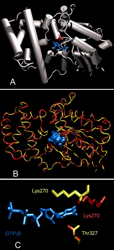
Figure 4. Computer generated model of the human α-cone transducin
A: Overall structure composition of GBT2_HUMAN (Genbank accession number P19087). The amino acid core of the protein is shown in gray, helical regions are displayed as cylinders. The GTP analog GTPγS is shown in blue and the deleted amino acid K270 is colored red. B: In this panel, the wild type (yellow) and the mutated protein structures (red) of GBT2_HUMAN were overlaid to show the overall structure integrity. The ligand is shown in blue. C: Three-dimensional representation of the essential amino acids involved in GTP binding in accordance with Figure 5D (wildtype is yellow; mutant is red).