![]() Figure 3 of
Sabah, Mol Vis 2004;
10:254-259.
Figure 3 of
Sabah, Mol Vis 2004;
10:254-259.
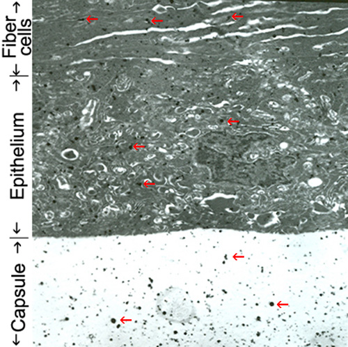
Figure 3. Localization of Alexa-albumin in the lens
Immunogold transmission electron microscopy of anterior, germinative region of lens after in vivo injection of Alexa-albumin. Labeling along the left margin of the figure delineates the boundaries between capsule, epithelium, and fiber cells. Red arrows designate representative enhanced gold particles that are uniformly distributed throughout the capsule, epithelium, and fiber cells.