![]() Figure 3 of
Karan, Mol Vis 2004;
10:248-253.
Figure 3 of
Karan, Mol Vis 2004;
10:248-253.
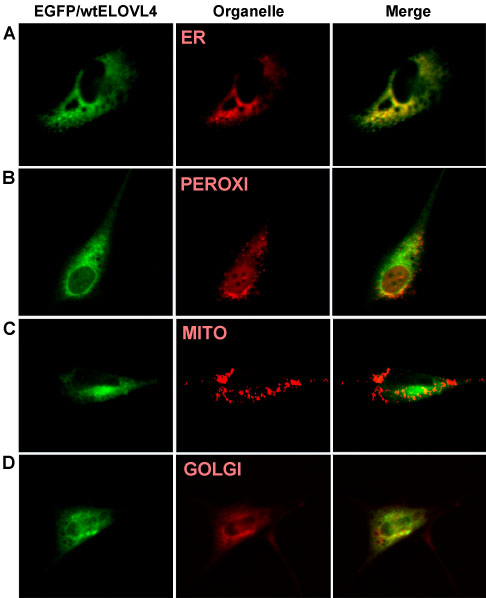
Figure 3. Intracellular localication of EGFP/wt ELOVL4 fusion protein
Localization of wtELOVL4-EGFP fusion protein with different organelle specific markers in NIH3T3 cells. The cells were imaged using confocal microscopy 24 h following co-transfection. Green images represent the expression of ELOVL4 and red images represent different organelles. Merged color image: superimposed image of green and red. A: Co-transfected cell with EGFP/wtELOVL4 and pDsRed2-ER (specific for Endoplasmic Reticulum). B: Co-transfected cell with EGFP/wtELOVL4 and pDsRed2-Peroxi (specific for peroxisomes). C: co-transfected cell with EGFP/wtELOVL4 and PDsRed2-Mito (specific for mitochondria). D: Cells were transfected with EGFP/wtELOVL4 and stained with Golgi specific marker BODYPY (red).