![]() Figure 3 of
Shentu, Mol Vis 2004;
10:233-239.
Figure 3 of
Shentu, Mol Vis 2004;
10:233-239.
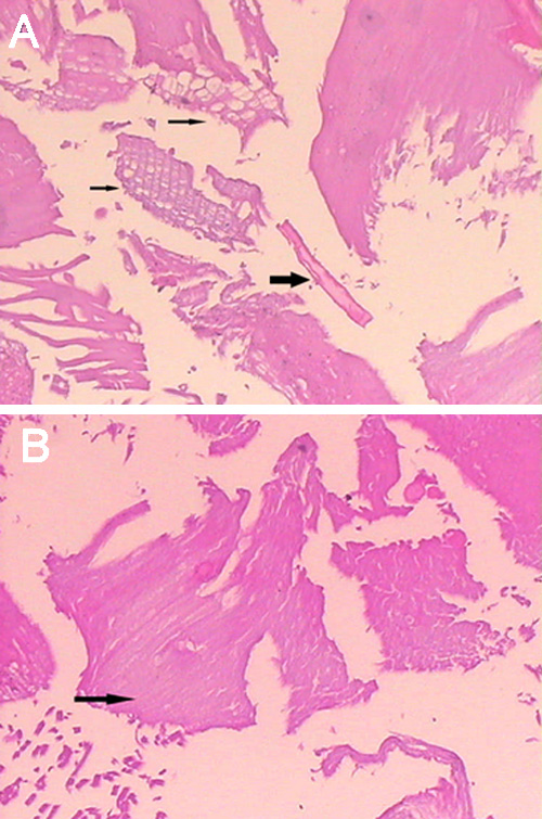
Figure 3. Pathology micrographs of removed lens tissue from affected individuals, by HE staining
There were focal degeneration alterations in the lens fiber cells. A: Reticular change (small arrow) and crystal precipitation (large arrow). B: Mucus-like degeneration (arrow).