![]() Figure 2 of
Kijas, Mol Vis 2004;
10:223-232.
Figure 2 of
Kijas, Mol Vis 2004;
10:223-232.
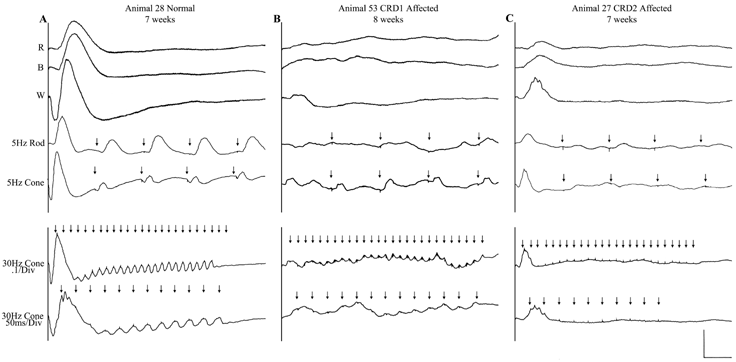
Figure 2. Electroretinograms from normal and cone-rod dystrophic dogs
Electroretinograms recorded from representative normal (column 1) and cone rod dystrophic (columns 2, 3) dogs. Each vertical panel presents the responses to a red flash (line 1); a blue flash (line 2); a white flash (line 3); 5 Hz low intensity white light flashes (line 4); and 30 Hz high intensity white light flicker (lines 5, 6): These elicit rod-specific (lines 2, 4), cone-specific (lines 5, 6, 7) and mixed rod-cone responses respectively. Animal 53 derives its disease from one founder (Animal 3, Figure 1) and Animal 27 from the other (Animal 1, Figure 1). Because these diseases are nonallelic they are termed crd1 and crd2, respectively. Although both cone and rod responses are markedly reduced in both affected dogs, the reduction in cone response amplitudes, relative to the loss of rod function, is far more severe than in previously described canine rod-cone degenerations and dysplasias. Residual cone function, at this age, is better preserved in animal 53 than Animal 27, a consistent albeit slight difference between CRD1 and CRD2 affected animals.