![]() Figure 4 of
Mackay, Mol Vis 2004;
10:155-162.
Figure 4 of
Mackay, Mol Vis 2004;
10:155-162.
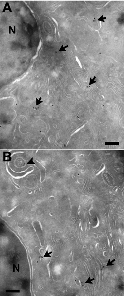
Figure 4. Ultrastructural localization of CRYGD in HLE B-3 cells
Cryo-immunoelectron microscopy of HLE B-3 cells expressing either wild type (A) or mutant (B) CRYGD showing that immuno-gold labeling (arrows in both panels) was primarily restricted to the cytoplasm. The arrowhead in B shows a double membrane structure surrounding immuno-gold particles. The letter N indicates the nucleus. The bar represents 1.5 μm. The magnification was x50,000.