![]() Figure 1 of
Rollin, Mol Vis 2004;
10:15-22.
Figure 1 of
Rollin, Mol Vis 2004;
10:15-22.
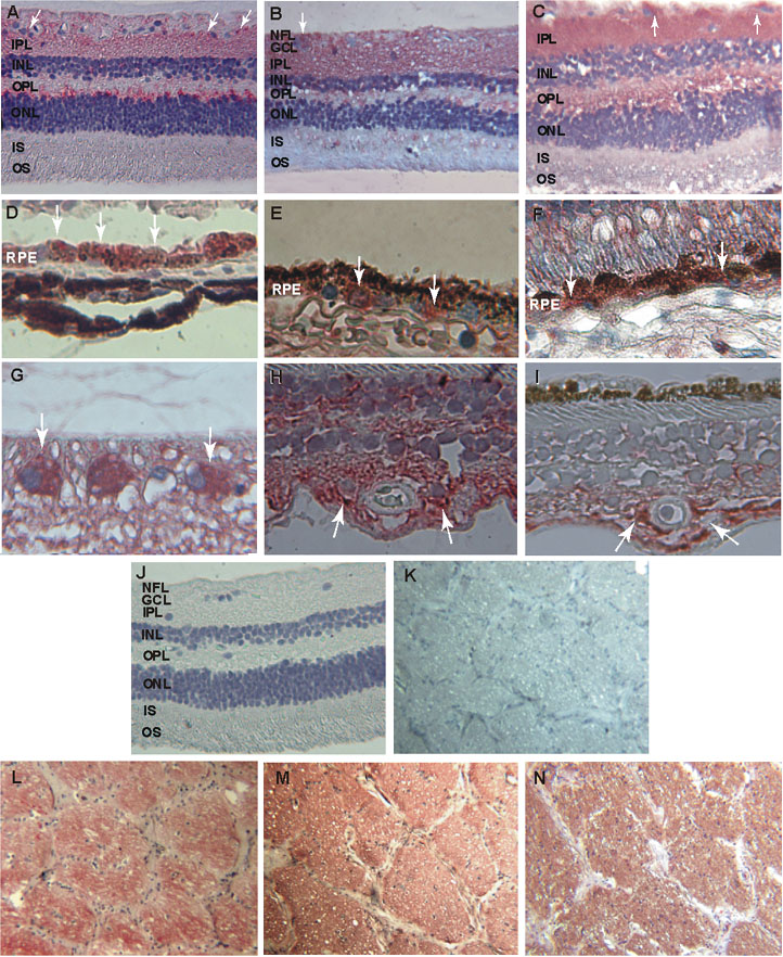
Figure 1. Localization of ANP, BNP, and CNP immunoreactivities
ANP, BNP, and CNP immunoreactivities were examined in human retina and the anterior portion of the optic nerve. Immunostaining appears in red. Positive ANP, BNP, and CNP (A-C, respectively, hematoxylin, x160) labeling can be seen in the inner and outer plexiform and nuclear layers of the retina. The cytoplasm of ganglion cells was also positive for the ANP, BNP, and CNP antibodies (arrows in A-C, respectively; BNP labeling is shown at higher magnification in G). RPE cells also showed intense labeling for ANP (D), BNP (E), and CNP (F; vertical arrows, hematoxylin, x400). ANP and GFAP immunoreactivity could be observed on astrocytes in adjacent sections (arrows, H, I, respectively, hematoxylin, x400) of the retina. Intense ANP, BNP, and CNP labeling (L-N, respectively, hematoxylin, x63) could also be seen in neural bundles of the anterior portion of optic nerves. Negative controls for the immunohistochemical detection of ANP, BNP, and CNP in the retina (J, hematoxylin, x160) and anterior portion of the optic nerve (K, hematoxylin, x63) were free of labeling. The meanings of the abbreviations used in this figure are: RPE (retinal pigment epithelium), OS (photoreceptor outer segments), IS (photoreceptor inner segments), ONL (outer nuclear layer), OPL (outer plexiform layer), INL (inner nuclear layer), IPL (inner plexiform layer), GCL (ganglion cells layer), NFL (nerve fiber layer).