![]() Figure 1 of
Yao, Mol Vis 2004;
10:138-143.
Figure 1 of
Yao, Mol Vis 2004;
10:138-143.
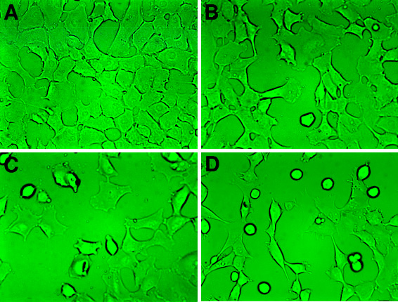
Figure 1. Phase-contrast microscopy analysis after microwave treatment
A: Control cells. B: Cells treated for 8 h with 0.50 mW/cm2 microwave radiation. Cells were comparable to the control. C: Cells treated for 8 h with 1.00 mW/cm2 microwave radiation. A few round, detached cells were visible in the microscopic field. D: Cells treated for 8 h with 2.00 mW/cm2 microwave radiation. A considerable number of round cells were present, and the density of adherent cells was diminished when compared with the control. The original magnification was 400x.