![]() Figure 3 of
Grus, Mol Vis 2004;
10:132-137.
Figure 3 of
Grus, Mol Vis 2004;
10:132-137.
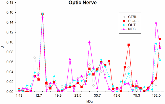
Figure 3. Antibody profiles against optic nerve antigens
The mean antigen-antibody reactivity of all groups were plotted vs. the corresponding molecular weight of the antigens. Complex staining patterns could be found in all groups. The NTG group revealed the highest difference from controls (p<0.002).