![]() Figure 1 of
Grus, Mol Vis 2004;
10:132-137.
Figure 1 of
Grus, Mol Vis 2004;
10:132-137.
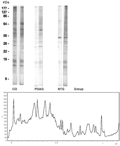
Figure 1. Photographs and densitographs of Western blots against optic nerve antigens
Top: Western blots of IgG autoantibody repertoires in subject sera against optic nerve antigens. There is a complex pattern of antibodies in all groups (CTRL, POAG, NTG). Each lane was incubated with subject serum (diluted 1:40) against bovine optic nerve over night. Secondary antibody, peroxidase-conjugated goat anti-human IgG (diluted 1:500), was applied for one hour. Bottom: Densitograph of a subject with primary open-angle glaucoma (POAG). In the densitograph scanner units ("U" represents optic density) were plotted against relative mobility (Rf-values) that correspond to the molecular weight of the antigenic tissue.