![]() Figure 5 of
Wang, Mol Vis 2004;
10:103-111.
Figure 5 of
Wang, Mol Vis 2004;
10:103-111.
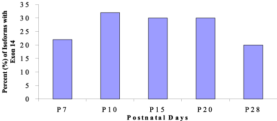
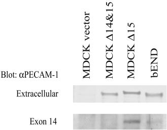
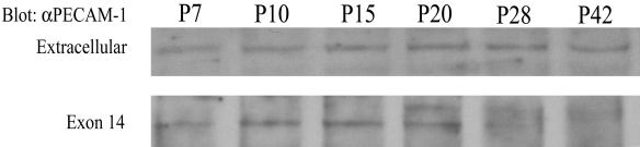
Figure 5. Western blot analysis of murine PECAM-1 isoforms
Cell lysates were prepared from MDCK cells transfected with the vector Δ15 or Δ14&15 PECAM-1 and brain endothelial (bEND) cells (A) or retinas from postnatal days P7, P10, P15, P20, P28, and P42 (B). Equal amounts of lysates (25 μg) from each sample were subjected to Western blot analysis as described in Methods. Blots were probed with an antibody to extracellular domain of murine PECAM-1, which recognizes all PECAM-1 isoforms. Same blots were also probed with an antibody to exon 14 of PECAM-1, which recognizes PECAM-1 isoforms with exon 14. Please note the exclusive presence of Δ15 PECAM-1 isoform in MDCK cells transfected with this isoform. The majority of PECAM-1 isoforms expressed in bEND cells lack exon 14 [3]. The percentage of murine PECAM-1 isoforms containing exon 14 relative to all the isoforms detected during vascularization of murine retina (derived from Table 2) are shown in C. This is very similar to the densitometric assessments of the band intensities in B. These experiments were repeated at least twice with similar results and a representative experiment is shown.
A:
B:
C: