![]() Figure 7 of
Wu, Mol Vis 2004;
10:93-102.
Figure 7 of
Wu, Mol Vis 2004;
10:93-102.
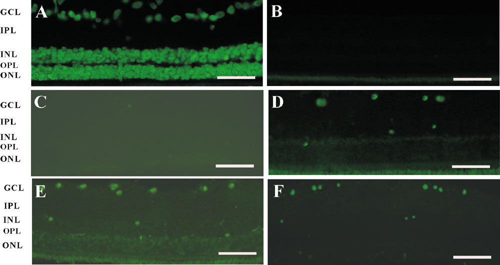
Figure 7. Apoptosis of RGCs detected by TUNEL staining 6 h after reperfusion
Three weeks after rAAV transduction, ischemia was induced for 60 min. Eyes were harvested 6 h after reperfusion, fixed and subjected to TUNEL analysis. A: A DNase treated sample, which stained positive for TUNEL staining and served as a positive control. B: Sample without the addition of the TdT enzyme showed no TUNEL staining and served as the negative control. C: Normal eye. D: rAAV-GDNF injected eye. E: rAAV-LacZ injected eye. F: Untreated eye. There were more TUNEL positive RGCs in rAAV-LacZ treated retina and in untreated retina than in rAAV-GDNF treated retina. Scale bar represents 50 μm. The outer nuclear layer (ONL), outer plexiform layer (OPL), inner nuclear layer (INL), inner plexiform layer (IPL), and the ganglion cell layer (GCL) are also labeled in the micrograph.