![]() Figure 6 of
Wu, Mol Vis 2004;
10:93-102.
Figure 6 of
Wu, Mol Vis 2004;
10:93-102.
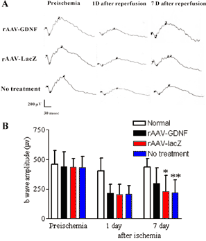
Figure 6. Effect of injury duration on ERG b-wave
A: Representative eletroretinographic recording. B: Mean b-wave amplitude before ischemia, and one and seven days after ischemia. There was no change of ERG in the normal control eyes throughout the experiment (n=8). One day after reperfusion, rAAV-GDNF treated eyes (n=28), rAAV-LacZ treated eyes (n=28), and untreated eyes (n=28) showed a reduction of b-wave amplitude, but there was no statistical difference between these groups. However, seven days after reperfusion, rAAV-GDNF treated eyes (n=26) showed significant recovery of b-wave amplitude than rAAV-LacZ treated eyes (n=26) and untreated eyes (n=26). Groups marked with an asterisk (*) were significantly different from the rAAV-GDNF transduced group (Wilcoxon signed-ranks test, p<0.05; Referent group: rAAV-GDNF transduced group, seven days after reperfusion). Groups marked with two asterisks (**) were significantly different from the rAAV-GDNF transduced group (Mann-Whitney test, p<0.05; Referent group: rAAV-GDNF transduced group, seven days after reperfusion). Error bars represent the standard deviation.