![]() Figure 2 of
Wu, Mol Vis 2004;
10:93-102.
Figure 2 of
Wu, Mol Vis 2004;
10:93-102.
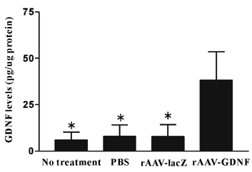
Figure 2. ELISA identifying retinal GDNF after rAAV-GDNF injection
Intravitreal injections of rAAV-GDNF (right eyes) and of rAAV-LacZ (left eyes) were performed (n=10). In a separate group of rats, intravitreal injection of PBS was performed in the right eye and no treatment was given in the left eye (n=7). Three weeks after rAAV transduction, tissue of the entire retina was harvested and processed for ELISA. Groups marked with an asterisk (*) were significantly different from the rAAV-GDNF transduced group (Wilcoxon signed-ranks test, p<0.05). Error bars represent the standard deviation.