![]() Figure 1 of
Wu, Mol Vis 2004;
10:93-102.
Figure 1 of
Wu, Mol Vis 2004;
10:93-102.
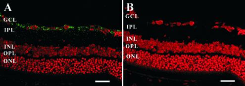
Figure 1. Immunohistochemistry of GDNF expression in rAAV-GDNF injected eyes
Rat eyes were transduced with either rAAV-GDNF (A) or rAAV-LacZ (B). Immunohistochemistry analysis of retinas using antibodies recognizing GDNF was performed three weeks after rAAV transduction. Cells possessing GDNF protein appear green (FITC), while nuclei stained with propidium iodide appear red. Scale bar represents 50 μm. The outer nuclear layer (ONL), outer plexiform layer (OPL), inner nuclear layer (INL), inner plexiform layer (IPL), and the ganglion cell layer (GCL) are labeled in the micrograph.