![]() Figure 3 of
Liao, Mol Vis 2004;
10:1038-1046.
Figure 3 of
Liao, Mol Vis 2004;
10:1038-1046.
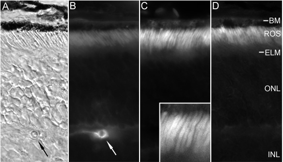
Figure 3. Immuofluorescent localization of albumin in the IRBP-/- mouse retina
A,B: Sections treated with FITC conjugated rabbit IgG directed against mouse albumin (A is the same section viewed by differential interference contrast microscopy; arrow indicates blood vessel). C: Control section that did not receive antibody. Insert is high magnification of outer segments. D: Section treated with antibody pre-adsorbed with purified mouse albumin. Bruch's membrane (BM), rod outer segments (ROS), external limiting membrane (ELM), outer nuclear layer (ONL) and inner nuclear layer (INL) are labeled.