![]() Figure 2 of
Liao, Mol Vis 2004;
10:1038-1046.
Figure 2 of
Liao, Mol Vis 2004;
10:1038-1046.
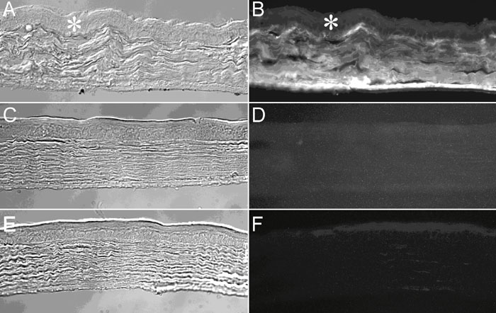
Figure 2. Localization of albumin in the IRBP+/+ mouse cornea stroma by immunofluorescence
A,B: Direct immunofluorescence using a rabbit antimouse albumin IgG conjugated with FITC. C,D: Control; section did not receive antibody. E-F: Antibody preadsorbed with mouse albumin. Asterisk identifies the corneal epithelium. A,C,E: Differential interference contrast microscopy; B,D,F: Fluorescence microscopy.